TPPP, golgi outposts and microtubules nucleation in oligodendrocytes
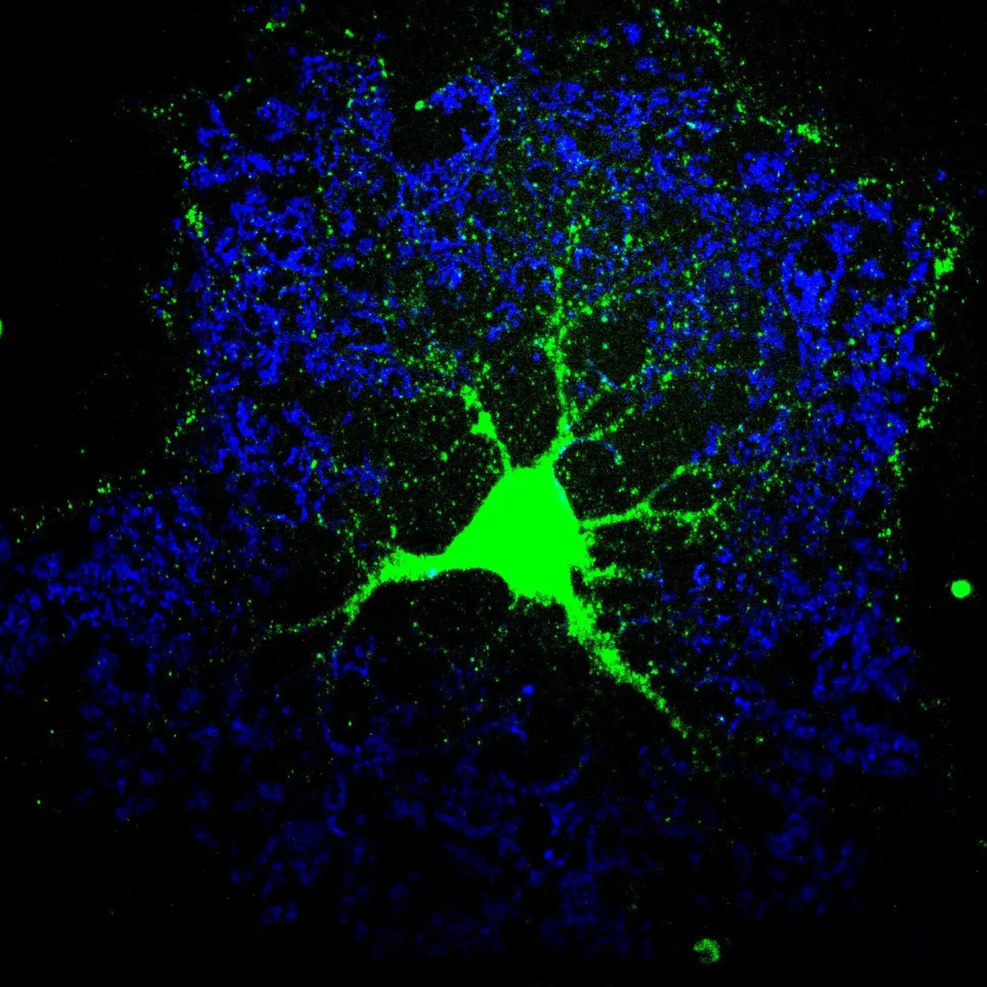
Green: Mbp mRNA
Blue: MBP protein
smFISH image of primary cultured rat oligodendrocyte
In our brains, electrically active neurons extend long processes called axons. These axons are insulated in fatty layers of membrane (much like rubber insulation around electrical wires) so that electrical signals can be sent quickly. The cells that make this insulation, called myelin, are oligodendrocytes.
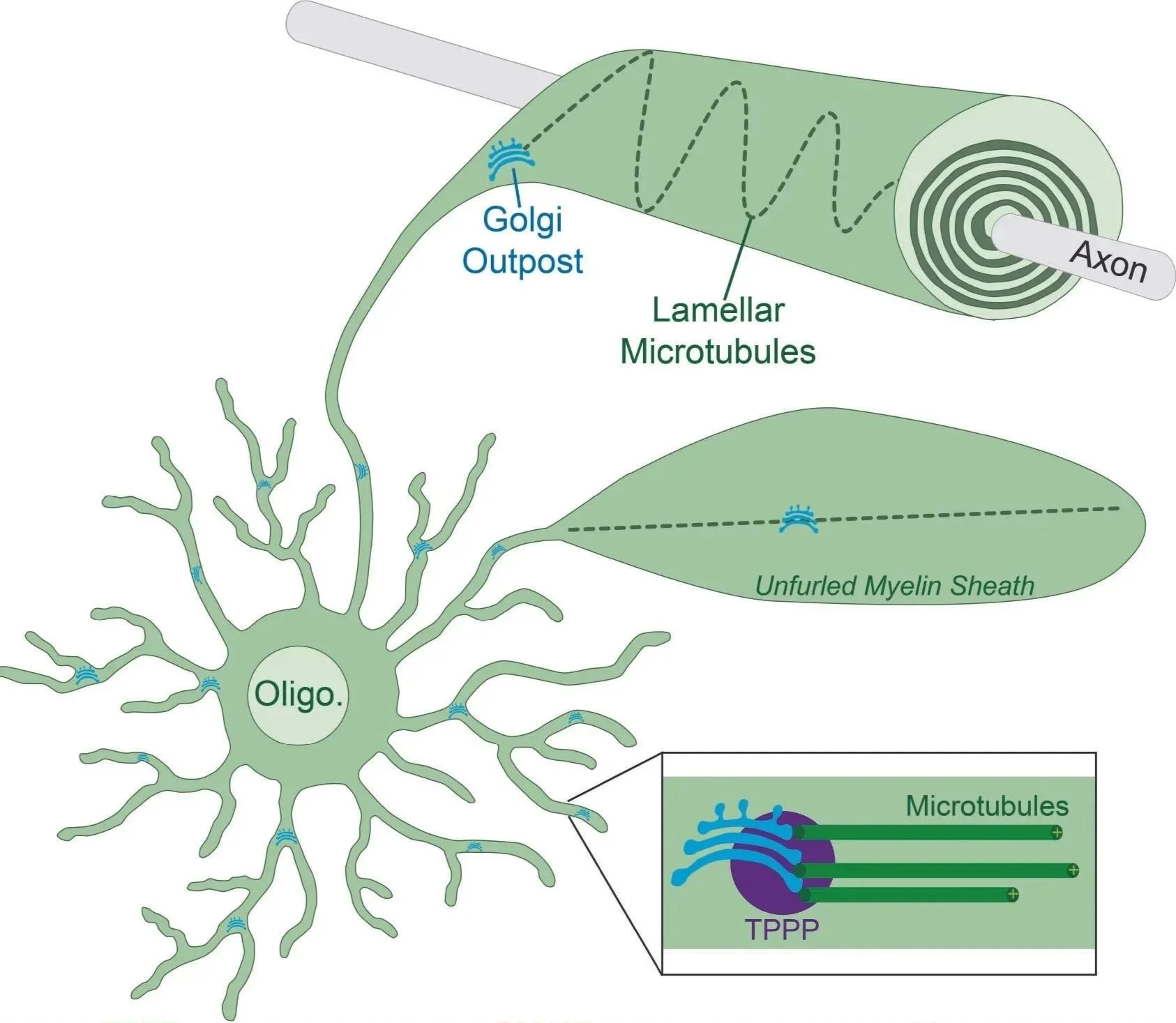
Oligodendrocytes are unique cells in the body that have extraordinary architecture. They are capable of making as many as 50 myelin sheaths per cell. Each cell extends branched processes that terminate in myelin sheaths that wrap about a dozen times around the axon. Microtubules, the train tracks of the cell, are found both inside these branches as well as inside myelin sheaths. Microtubules are important for the structure of the myelin sheath and for long-distance transport of cargos (organelles, mRNAs, proteins).
Due to the complex architecture of oligodendrocytes, microtubules are formed at distances far from the cell body. Our lab discovered that they do this using Golgi outposts and the associated protein TPPP (tubulin polymerization promoting protein).
TPPP can aggregate in neurological diseases, including MSA (multiple system atrophy) and Parkinson’s disease. We recently discovered that this is due to TPPP’s property as a liquid condensate. Much like oil in water, TPPP proteins are able to phase separate by interact with other TPPP proteins and partner proteins, like tubulin (the building blocks of microtubules).
mbp mrna transport in oligodendrocytes
The most abundant mRNA in oligodendrocytes is MBP (myelin basic protein). MBP functions within myelin sheaths to bring adjacent membranes together (much like a molecular glue) in order to form more efficient insulating layer. This process is called compaction and effectively squeezes out cytoplasm from the myelin sheath, much like squeezing toothpaste out of a tube.
As a result of this function, MBP mislocalization within the cell body can lead to adhesion between organelles and plasma membrane, for example. Thus, proper MBP localization is highly regulated. Mbp mRNA is transported to the myelin sheath, then it is locally translated. We are currently combining many techniques to understand the mechanism of how this happens.
Using biochemistry, we are solving the structure of Mbp mRNA using an approach called DMS-MaPseq. Using primary oligodendrocyte cultures, we can look at mRNA localization using smFISH and live-cell imaging. Using transgenic mouse lines, we can ask what the effects of perturbing these processes does to the brain and whether there are any behavioral effects. For example, by replacing the endogenous 3’ UTR region of Mbp, we generated a mouse that presents in the first few weeks of life with tremors.
MBP translation is hindered in myelinating diseases like MS (multiple sclerosis) and childhood developmental diseases like leukodystrophies. Thus, a basic understanding of the cell biology underlying MBP expression is critical for understanding disease.
astrocyte cytoskeleton
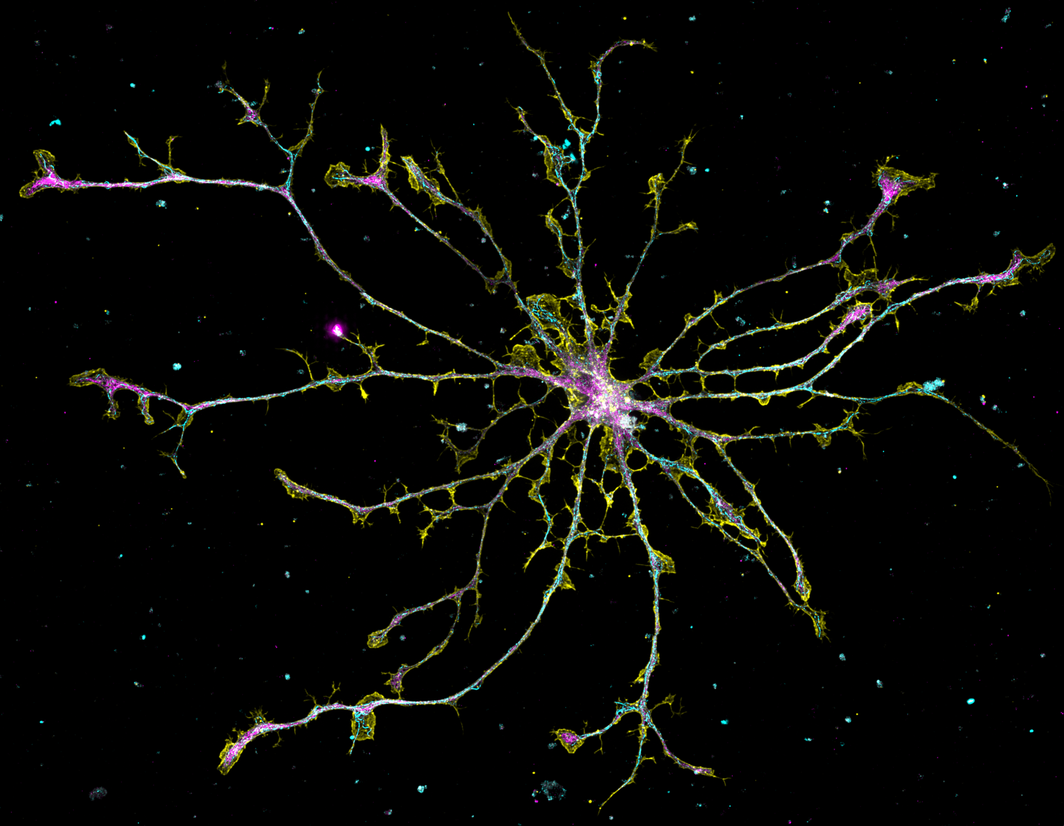
Cyan: Microtubules
Yellow: actin
Magenta: GFAP
Astrocytes are glial cells in the brain that maintain synapses and form the blood-brain barrier. They do so by extending processes that contact neurons and blood vessels. A new project in our lab seeks to understand how 3 classes of cytoskeleton (“cell skeleton”) - microtubules, actin, intermediate filaments - build the astrocyte.
These basic cell biology questions are important to understand astrocyte function and why this is important in the brain. Cytoskeleton establish cellular tracks that allow cargos to move around in cells. For example, we recently found that microtubules are organized in a uniform orientation with plus ends directed outwardly.
In addition, mutations in the intermediate filament gene GFAP (glial fibrillary acidic protein) can lead to Alexander’s disease, a leukodystrophy or neurodevelopmental disorder affecting children that is characterized by seizures and autism-like symptoms.
astrocyte cell biology in the blood-brain barrier (BBB)
Vascular dementia is second most common cause of dementia (behind Alzheimer’s). A new project studies this enigmatic disease at the biochemical, cellular, and animal levels. At the animal level, we generated a mouse model for a rare genetic form of vascular dementia. At the cellular level, evidence indicates that the blood-brain barrier, which protects the brain from pathogens, fails in vascular dementia. Thus, we have developed methods to culture endothelial cells from brain blood vessels together with astrocytes to mimic the blood-brain barrier in a dish. Finally, we will perform biochemistry experiments to determine the partner proteins that are involved in the pathology of this disease.